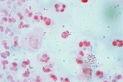
Corynebacterium diphtheriae
Gram-positive bacterium causing diphtheria, toxigenic via bacteriophage.

Corynebacterium diphtheriae is a Gram-positive pathogenic bacterium that causes diphtheria. The bacterium primarily infects humans, causing respiratory diphtheria, and its virulence is enhanced when infected by a bacteriophage carrying a toxin gene.
- discovered_by
- Edwin Klebs and Friedrich Löffler
- type
- Gram-positive bacterium
- disease_caused
- Diphtheria
- subspecies
- C. d. mitis, C. d. intermedius, C. d. gravis, C. d. belfanti
- toxin_mechanism
- Inactivates elongation factor EF-2 via ADP-ribosylation
Lore & Background
Corynebacterium diphtheriae is a rod-shaped, Gram-positive, non spore-forming, nonmotile bacterium that exclusively infects humans, though rare cases have been found in animals. The bacterium causes diphtheria by adhering to mucosal layers, primarily in the respiratory tract, and releasing an exotoxin that inhibits protein synthesis in host cells. The diphtheria toxin is encoded by a corynephage integrated into the bacterial chromosome. Toxin production is controlled by iron concentration; low iron levels promote toxin production, while high iron levels shut it down via an aporepressor. Four subspecies are recognized, differing in colonial morphology and biochemical properties, and all may be toxigenic or nontoxigenic. Identification involves Gram stain showing pleomorphic organisms resembling Chinese letters, and stains like Albert's and Ponder's to demonstrate metachromatic granules. Enrichment media such as Löffler's medium and tellurite agar are used, with C. diphtheriae producing brown colonies or black halos. The bacterium produces catalase but not urease, differentiating it from related species.
Reader's Guide
The bacterium's virulence depends on infection by a bacteriophage carrying the toxin gene, which inactivates elongation factor EF-2, leading to pseudomembrane formation in the throat and potential systemic damage to kidneys, nerves, and heart. The incidence of diphtheria has decreased sharply in vaccinated populations, but outbreaks continue in developing countries with low vaccination coverage. Cutaneous diphtheria, prevalent in tropical climates, may contribute to outbreaks. The bacterium is part of the C. diphtheriae species complex, which includes zoonotic pathogens like C. ulcerans and C. pseudotuberculosis that can also produce diphtheria toxin. Strain subtyping for outbreak tracking remains limited due to lack of publicly available resources. The DTaP vaccine effectively prevents the disease and is mandatory in the United States for public education and some professions.
Did You Know?
- The diphtheria toxin gene is carried by a bacteriophage integrated into the bacterial chromosome.
- Four subspecies are recognized: C. d. mitis, C. d. intermedius, C. d. gravis, and C. d. belfanti.
- The bacterium produces catalase but not urease, differentiating it from Corynebacterium ulcerans.
Discovery, Nomenclature & Taxonomic Placement
Their joint discovery earned the bacterium its enduring alternative name, the Klebs–Löffler bacillus, a nod to the collaborative spirit of nineteenth-century microbiology. The organism is nonmotile and does not form spores, traits that help set it apart from other bacterial threats of the era. Taxonomically, C. diphtheriae sits within a broader species complex that also encompasses C. belfantii, C. pseudotuberculosis, C. rouxii, C. silvaticum, C. ulcerans, and C. ramonii, a grouping that reflects shared evolutionary lineage. Within the species itself, four subspecies are recognized—mitis, intermedius, gravis, and belfanti—each showing subtle differences in colonial morphology and in the capacity to metabolize particular nutrients. Every one of these subspecies can exist in either a toxigenic or a non-toxigenic form, a distinction that ultimately determines whether the bacterium poses a lethal threat or remains a relatively benign colonizer.
The Phage-Borne Toxin and Its Iron-Gated Regulation
The true lethality of C. diphtheriae does not reside in the bacterium itself but in a viral passenger. A bacteriophage known as a corynephage integrates its genetic cargo into the bacterial chromosome during the lysogenic phase, depositing a Tox gene that encodes the diphtheria toxin. Without this phage infection, the strain is essentially harmless. Once expressed, the toxin wreaks havoc by inactivating elongation factor EF-2, a protein essential for host-cell translation, which halts protein synthesis and produces the characteristic pharyngitis and the thick grey pseudomembrane lining the throat. Production of the toxin is tightly regulated by iron: at low iron concentrations the repressor stays inactive and toxin is made, whereas at high iron levels the metal binds an aporepressor on the beta bacteriophage, effectively switching off the Tox gene. In the laboratory, Elek's test is the standard method for confirming whether a given isolate is toxigenic. Notably, certain zoonotic relatives such as C. ulcerans and C. pseudotuberculosis can also harbor the same phage and produce an identical toxin, underscoring that the danger lies in the gene rather than the host species.
Clinical Course, Transmission & Human Exclusivity
C. diphtheriae is a pathogen almost exclusively of humans, with only two documented cases in dogs and two in horses, all of them toxigenic. The bacterium typically enters through the nose, tonsils, or throat and adheres to mucosal surfaces before infiltrating deeper tissue layers. The hallmark of respiratory diphtheria is a thick, grey pseudomembrane that accumulates in the nasopharyngeal region, progressively obstructing breathing and swallowing. Early symptoms include sore throat, fever, weakness, and swollen cervical glands, but the danger escalates rapidly if the toxin enters the bloodstream, where its proteolytic activity can devastate the heart, kidneys, and peripheral nerves. In the most extreme cases, patients face suffocation or partial paralysis. Transmission occurs primarily through respiratory droplets generated by coughing or sneezing, though contact with open skin lesions or contaminated surfaces is a less common route. A critical public-health fact is that an individual remains contagious for at least two weeks after visible symptoms resolve, and in some instances for as long as a full month, making contact tracing and isolation essential.
Vaccination, Global Inequity & the Tropical Reservoir
For much of the nineteenth and early twentieth centuries, diphtheria was a leading killer of children, and the prospect of a vaccine transformed the disease's trajectory. Yet the story is far from over. In developing countries and tropical regions where a large share of the population lacks access to routine immunization, outbreaks still erupt with devastating frequency. Cutaneous diphtheria—skin lesions harboring toxigenic strains—is particularly prevalent in warm, tropical climates and can serve as a reservoir that seeds new respiratory outbreaks. The groups most vulnerable today are unvaccinated children, poorly immunized adults, and immunocompromised individuals. The persistence of the disease in under-resourced settings is a stark reminder that the bacterium itself has not changed; only the uneven distribution of the tools to neutralize it has.
Frequently Asked Questions
What are Corynebacterium diphtheriae's powers/role?
Its signature weapon is the diphtheria toxin, a protein it only produces after a specific bacteriophage inserts a toxin gene into its genome. That toxin kills host cells by ADP-ribosylating elongation factor EF-2, which halts protein synthesis and can cause tissue necrosis in the throat or elsewhere.
How does Corynebacterium diphtheriae's story end?
Clinically, the infection is managed with diphtheria antitoxin plus antibiotics such as penicillin or erythromycin, which neutralize circulating toxin and eliminate the bacteria. Widespread toxoid vaccination has made the disease rare in most developed nations, though sporadic outbreaks still occur in underserved regions.
Why is Corynebacterium diphtheriae important?
It is the textbook example of lysogenic conversion, showing how a harmless phage can turn a mild organism into a deadly pathogen through horizontal gene transfer of the toxin gene. It also remains a WHO-listed notifiable disease and a key model for studying phage-bacteria interactions in microbiology.
What subspecies does Corynebacterium diphtheriae have?
It is divided into four subspecies—mitis, intermedius, gravis, and belfanti—each with distinct colony morphology and virulence tendencies. The gravis subspecies is generally linked to the most severe disease, while belfanti often causes milder or asymptomatic carriage.
More in Pathogenic Bacteria 1-20
Spotted an error? Know more?
This is a living reference — every entry is fact-audited, and reader corrections feed straight into our audit queue. Suggest an edit · See this site's audit record